
| name | Amanita hongoi |
| name status | nomen acceptum |
| author | Bas |
| english name | "Hongo's Lepidella" |
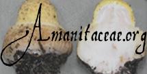
| images |
.") 1. Amanita hongoi, Japan (drawn by C. Bas from watercolor of T. Hongo).  2. Amanita hongoi, China. |
| intro |
The following combines the description of Bas (1969) with data collected by Z. L. Yang. |
| cap |
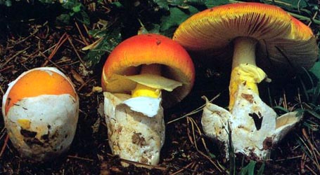
The cap of A. hongoi is 70 - 170 mm wide, at first hemispherical, then convex to plane, finally somewhat depressed at the center, rather fleshy, white to brownish to yellowish to pale yellowish brown, dry, with a nonsulcate, appendiculate margin. The cap is regularly decorated with medium-sized, brownish, conical warts of volval material that are pyramidal to subpyramidal to subconic, diminishing in size towards the margin. The warts are 1 - 3 mm high, brownish, dirty yellow to pale yellow-brown. The margin of the cap is nonstriate and appendiculate. |
| gills |
The gills are crowded, free, rather broad, and white to cream. The short gills are rounded-truncate (to attenuate?). |
| stem |
The stem is 80 - 150 × 5 - 45 mm, solid, white to dirty white, becoming brownish with age, flocculose-squamulose, slightly tapering upward, and annulate. The stem is covered with white to brownish squamules. The lower half has many close circles of very minute, brownish, conical to pustule-like warts. The annulus is subapical, white, and fugacious. |
| spores |
The spores measure (7.0-) 7.5 - 9.5 (-11.0) × (6.0-) 6.5 - 8.0 (-9.5) µm and are amyloid and globose to broadly ellipsoid. Clamps are not found at bases of basidia. |
| discussion |
This species was originally described from Japan (Honshu) where it occured in association with fagaceous trees (such as oak) in coniferous-deciduous forest. The species is now also known to occur in China, Korea, Japan, and peninsular southeast Asia. Bas placed A. hongoi in his stirps Perpasta (see
A. perpasta Corner & Bas). |
| brief editors | RET |
| name | Amanita hongoi | ||||||||
| author | Bas. 1969. Persoonia 5: 410, figs. 130-132. | ||||||||
| name status | nomen acceptum | ||||||||
| english name | "Hongo's Lepidella" | ||||||||
| MycoBank nos. | 308559 | ||||||||
| GenBank nos. |
Due to delays in data processing at GenBank, some accession numbers may lead to unreleased (pending) pages.
These pages will eventually be made live, so try again later.
| ||||||||
| holotypes | L | ||||||||
| revisions | Z. L. Yang and Y. Doi. 1999. Bull. Natl. Sci. Mus. Tokyo B 25(3): 118, fig. 15. | ||||||||
| selected illustrations |
Imazeki and Hongo. 1987. Color. Illus. Mushr. Japan 1: 133, pl. 33 (fig. 230). Imazeki and Hongo. 1965. Color. Illus. Mushr. Japan 2: 43-44, pl. 13 (fig. 80). [As "Amanita echinocephala."] | ||||||||
| intro |
The following text may make multiple use of each data field. The field may contain magenta text presenting data from a type study and/or revision of other original material cited in the protolog of the present taxon. Macroscopic descriptions in magenta are a combination of data from the protolog and additional observations made on the exiccata during revision of the cited original material. The same field may also contain black text, which is data from a revision of the present taxon (including non-type material and/or material not cited in the protolog). Paragraphs of black text will be labeled if further subdivision of this text is appropriate. Olive text indicates a specimen that has not been thoroughly examined (for example, for microscopic details) and marks other places in the text where data is missing or uncertain. The following material is derived from the protolog of the present taxon, and the original research of Drs. Z. L. Yang and Y. Doi. NOTE: Spore measurements from papers by Z. L. Yang use his "Times New Roman" face for "Q" and "Q'"—respectively, " from protolog Basidiomes large to very large, thickset. | ||||||||
| pileus | from protolog: 150 - 170 mm wide,white to brownish, at first hemispheric, then convex to planar, finally somewhat depressed in disc, dry; context fleshy, rather soft, white, unchanging; margin non-sulcate, appendiculate; universal veil as regularly distributed warts, medium-sized, brownish, conical, ca. 2 - 3 × 2 - 4 mm, diminishing in size toward margin. | ||||||||
| lamellae | from protolog: free, crowded, white, up to 15 mm wide, with minutely flocculose edge; lamellulae rounded truncated (to attenuate?). | ||||||||
| stipe | from protolog: 110 - 130 × 20 - 30 mm (width measured at apex), white, becoming brownish with age; bulb as clavate base, 40 - 45 mm wide, context solid, white, rather soft, unchanging; partial veil apical, membranous, pendent, thick, 25 - 30 mm wide, cream, striate above, floccose below, bearing warts of universal veil on underside at edge (per figure); universal veil distributed on lower half of stipe in many close circles of very minute warts, brownish, conical to pustule-like. | ||||||||
| odor/taste | from protolog: Odor agreeable. Taste mild. | ||||||||
| macrochemical tests |
none recorded. | ||||||||
| pileipellis | from protolog: thin, slightly gelatinized at shiny dark spots between warts; filamentous hyphae 2 - 10 μm wide, interwoven; vascular hyphae plentiful. [Note: Original text seems to imply that the dimensions of the vascular hyphae are included in the dimensions given here for the filamentous hyphae.—ed.] | ||||||||
| pileus context | not described. | ||||||||
| lamella trama | from protolog: difficult to analyze in available material. | ||||||||
| subhymenium | from protolog: cellular. | ||||||||
| basidia | from protolog: 45 - 55 × 11 - 13 μm, mostly 4-, but infrequently 2- or 1-sterigmate, many with refractive content; clamps lacking. | ||||||||
| universal veil | from protolog: On pileus, upper part: yellow in alkaline solution, with elements having more or less erect-parallel orientation; filamentous hyphae inconspicuous, branching, 3 - 8 μm wide; inflated cells predominantly broadly ellipsoid, 15 - 45 × 10 - 35 μm, small, yellowish, in irregular terminal chains; vascular hyphae scattered, yellow. On pileus, lower part: inflated cells fewer and smaller; vascular hyphae 3 - 20 μm wide, abundant, branching, yellowish. On pileus, peripheral part of base: on surface here and there bearing patches of amorphous yellow material ("excreted by oleiferous hyphae?"); vascular hyphae comprising entire tissue, yellow, 3 - 8 μm wide. On stipe base: filamentous hyphae branching, sometimes with yellow refractive contents; inflated cells small, abundant. [Note: The term "oleiferous" has now been replaced by "vascular" because there is no evidence of oil in Amanita hyphae (per conversations with Dr. Bas).—ed.] | ||||||||
| stipe context | from protolog: longitudinally acrophysalidic; filamentous hyphae "rather broad." | ||||||||
| partial veil | not described. | ||||||||
| lamella edge tissue | from protolog: scanty; inflated cells small, up to 20 μm wide, mostly in short chains. | ||||||||
| basidiospores |
from protolog: [20/1/1] (7.0-) 8.0 - 10.0 (-11.5) × 7.0 - 9.0 (-10.0) μm, (Q = 1.0 - 1.20; Q = 1.10), colorless, smooth, thin-walled, amyloid, globose to broadly ellipsoid, often more or less obpyriform; contents guttulae or homogeneous and refractive. Yang and Doi (1999): [30/1/1] (7.5-) 8.0 - 10.0 × (6.5-) 7.0 - 8.5 (-9.0) μm, ( | ||||||||
| ecology | Solitary to subgregarious. Japan: In Quercus forests (holotype under Q. serrata Thunb.) or in coniferous and deciduous forest. | ||||||||
| material examined |
from protolog: JAPAN: HONSHU—Ôtsu City - ca. Sotohata, 25.i.x1958 T. Hongo 1881 (holotype, L). Yang and Doi (1999): JAPAN: HONSHU—Tokyo Metropolis (Pref.) - Oume City, Kurosawa 3-chôme, right-side ridge of Kurosawa-gawa R., 25.ix.1997 Y. Doi s.n. (TNS F-237946). | ||||||||
| discussion |
from protolog: "I have not examined the part of Hongo 1881 in Dr. Hongo's private herbarium. "The tissues of the type are rather difficult to study. It was possible to analyze the tissue of the warts on the cap, but impossible to make a good illustration of it. "Undoubtedly Amanita hongoi from Japan is closely related to A. perpasta from Malaya. It has the same habit, the same type of ring, similar spores, rather similar conical warts on the cap, etc. The coloured spots around the base of the warts, which are such a remarkable feature of A. perpasta, are also present in A. hongoi, though to a much lesser degree. "Amanita perpasta, however, has considerably coarser warts on the cap; in a young stage these already have a subferrugineous-brown tip and a whitish to somewhat brownish base, whereas the smaller, entirely brownish warts in A. hongoi are situated directly on the surface of the cap. In addition to this there are several less conspicuous differences between the two species: (i) In alkaline solution the tissue of the warts of A. hongoi is yellow because of the deep yellow oleiferous hyphae, especially abundant in the base of the warts, and the yellowish inflated cells; the tissue of the warts of A. perpasta is coloured only near the surface but is nearly colourless in the base of the warts, where only scattered, rather narrow oleiferous hyphae are present. (ii) The spores of A. hongoi are slightly larger than those of A. perpasta (8 - 10 × 7 - 9 μm against 7 - 8.5 × 6.5 - 8 μm). (iii) The ring of A. hongoi is floccose at the underside and not coarsely verrucose like in A. perpasta. [Note: The term "oleiferous" is discussed in the "universal veil" data field, above.—ed.] The following figure provides a sporograph based comparison of the two species discussed above: "In several respects A hongoi resembles A. solitaria...from Europe. That species, however, is different in that it has clamps, larger inflated cells in the warts on the cap, larger, more elongate spores, greenish-yellow tinged gills, etc." | ||||||||
| citations | —R. E. Tulloss | ||||||||
| editors | RET | ||||||||
Information to support the viewer in reading the content of "technical" tabs can be found here.
| name | Amanita hongoi |
| name status | nomen acceptum |
| author | Bas |
| english name | "Hongo's Lepidella" |
| images |
1. Amanita hongoi, Japan (drawn by C. Bas from watercolor of T. Hongo). 2. Amanita hongoi, China. |
| photo | Zhu L. Yang - (2) China. |
| drawing |
C. Bas - (1) reproduced by courtesy of Persoonia, Leiden, the Netherlands from Bas (1969) based on watercolor of Dr. T. Hongo (Japan). |
| name | Amanita hongoi |
| bottom links |
[ Section Lepidella page. ] [ Amanita Studies home. ] [ Keys & Checklists ] |
| name | Amanita hongoi |
| bottom links |
[ Section Lepidella page. ] [ Amanita Studies home. ] [ Keys & Checklists ] |
Each spore data set is intended to comprise a set of measurements from a single specimen made by a single observer; and explanations prepared for this site talk about specimen-observer pairs associated with each data set. Combining more data into a single data set is non-optimal because it obscures observer differences (which may be valuable for instructional purposes, for example) and may obscure instances in which a single collection inadvertently contains a mixture of taxa.


Text and User-Generated Sporographs are published under the Creative Commons License.

In the case of a taxon page, image credits are on the 'image' tab.